Panáková Lab
Profile
The central idea of our research is to understand how physical cues interact with major signaling pathways during development and in disease.
We use zebrafish as a model system.
Zebrafish is particularly suited to study embryonic development due to its short generation time, small size, genetic and chemical tractability, transparency of the embryos and external fertilization that allows for the in vivo observation and manipulation of early developmental events.
We focus on cardiovascular development.
Heart is the first functional organ in developing vertebrate embryo. The zebrafish heart begins to synchronously contract already at 24 hours post fertilization (hpf) and retains its epithelial characteristics up to at least 90 hpf. The cardiomyocytes are coupled together to form electrochemical syncytium that leads to spontaneous and synchronized electrical activity. These are the assets that provide unique settings for studying the interaction between developmental pathways and electrochemical cues.
We use integrative approaches.
From classical genetic tools to high speed optical mapping techniques, we use a number of different approaches to answer our questions.
Team
Team Members
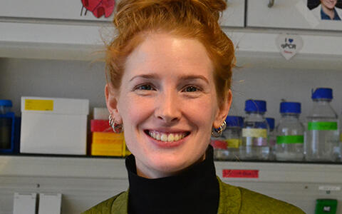
Daniela Panáková, Group Leader
I finished my PhD with Suzanne Eaton at Max Planck Institute of Molecular Cell Biology and Genetics in Dresden, Germany in 2005. After a short postdoctoral stay in her lab, I moved to Boston, MA and worked with Calum MacRae at Harvard Medical School/Brigham and Women's Hospital until 2011. I established my independent research group at Max Delbrück Center for Molecular Medicine in July, 2011 after obtaining Helmholtz Young Investigator Program Grant. I am interested in investigating the interactions between physiology and signaling pathways throughout development and in applying the obtained knowledge to understand the mechanisms underlying common disease states.
Alexander Meyer, Research Assistant
I graduated from the Beuth University of Applied Sciences in Berlin with major in Biotechnology. I joined the lab in August 2011 as a Research Assistant. Besides cloning, ordering or regular lab chores, I also work on a collaborative project with Matt Poy lab, in which we investigate the role of cell adhesion molecule CADM1 in pancreatic development.
Mariana Guedes Simoes, Post doc
Anne Merks, Postdoc
Laura Bartolini, PhD student
I finished my Master's Degree at University of Bologna in spring 2012 and soon after moved to MDC, Berlin. The main idea of my PhD project is to elucidate the role of electrochemical signals during angiogenesis. Particularly, I am interested how calcium fluxes regulate the tip cell behavior during vessel outgrowth using well-established zebrafish model.
Mai Phan, PhD Student
Nicola Müller, PhD Student
Kevin Manuel Mendez Acevedo, PhD Student
Sara Lelek, , PhD Student
Past members
- Laura Lleres Forero, Postdoc
- Tareck Rharass, Postdoc
- Motahareh Moghtadaei, Post doc
- Marie Swinarski, PhD student
- Kitti Csályi, PhD student
- Niels van der Maaden, Intern
- Zahir Shivji, Intern
- Marek Svoboda, Intern
- Ines Uhlenhut, Bachelor student
Research
Research Focus
Physiological cues that include electrical and ionic impulses or metabolic intermediates have been long implicated in the orchestration of organ development during early embryonic patterning. While much is known about genetically encoded developmental pathways, the mechanisms by which these small molecules interact during embryogenesis are poorly understood. We have developed technologies to explore the role of electrochemical and other physiologic stimuli during development in the zebrafish. Recently, we have demonstrated that Wnt11 non-canonical signaling, a major developmental pathway regulating tissue morphogenesis and organ formation, patterns intercellular electrical coupling in the myocardial epithelium through effects on transmembrane calcium conductance mediated via the L-type calcium channel. This finding offers a new entry point into an aspect of Wnt non-canonical signaling that has been proven difficult to explore. We focus on studying the Wnt/calcium pathway in the context of the developing embryo utilizing various techniques ranging from electrophysiology to developmental biology. Our immediate interest is to describe the molecular mechanisms that lead to the Wnt-mediated attenuation of L-type calcium channels, to characterize how non-canonical Wnt signals compartmentalize and affect different calcium domains in various tissues, and to determine how electrochemical signals modulated by Wnt11 regulate cardiac morphogenesis and function.
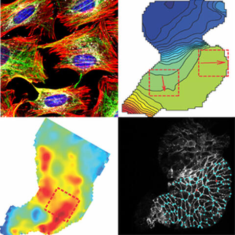
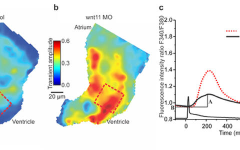
Wnt11 regulates Ca2+ transient amplitudes in cardiomyocytes: a,b Colour maps of Ca2+ transient amplitudes from control hearts (a) and Wnt11 morphants (b). Colour code depicts Ca2+ transient amplitudes in fluorescence ratio units (F340/F380). Squares indicate automated ROIs for measurements averaged in c. (c) Averaged Ca2+ transients from ROIs in a,b.
Research Projects
Project 1: Molecular bases of interactions between Wnt non-canonical pathway and L-type Ca2+ channel.
While we have shown that Wnt11 non-canonical signaling modulates the electrical coupling of cardiomyocytes via negative regulation of the L-type Ca2+ channel (LTCC) conductance, there are many questions that remain to be answered. How does Wnt11 signaling modulate LTCC function? Is the interaction between Wnt11 and LTCC direct or indirect? What known downstream components of Wnt11 signaling pathways are involved in the LTCC attenuation? In order to define the molecular mechanism by which Wnt11 regulates LTCC at a subcellular resolution, we have moved from zebrafish embryonic hearts to cell-based systems. This allows us to perform not only immunological experiments, but also more importantly detailed biochemistry.
Project 2: Characterization of the components of the Wnt non-canonical transduction pathway involved in the L-type Ca2+ channel attenuation.
The specificity and a wide range of diverse biological effects of Wnt signaling are mediated via binding and affinity of distinct Wnts to their corresponding receptors, Frizzleds (Fzd). It has been shown that Wnt11 interacts with Fzd7 genetically as well as physically. A crucial step in Wnt signaling is the translocation of Dishevelled (Dsh) from the cytoplasm to the plasma membrane, relaying the Wnt signal to various downstream effectors. Dsh has been implicated so far in all known aspects of Wnt signaling, canonical and non-canonical. We aim to define the requirement of the Wnt transduction machinery in the process of attenuation of L-type Ca2+ channel.
Project 3: Role of Wnt11/Ca2+ signaling in cardiac development.
We have demonstrated that Wnt11 patterns electrical coupling through regulation of LTCC conductance. However, we do not know what cellular responses precede this patterning. It is feasible to envisage the development of electrical coupling as a process of junction formation. Indeed, Wnt non-canonical signaling has been implicated in regulating cell adhesion and junctional remodeling in many cell biological and developmental contexts. Hence, we have begun addressing the question whether Wnt/calcium signaling is involved in junction formation.
Project 4: Quantitative analysis of Ca2+ compartmentalization by non-canonical Wnt-dependent mechanisms.
To further understand the role of Wnt11 signaling in regulating Ca2+ fluxes, we decided to take a systematic approach. We are taking advantage of the ratiometric high-resolution Ca2+ imaging with Fura-2, that we have developed, to perform quantitative analysis of Ca2+ fluxes in both excitable and non-excitable tissues. The final aim is to create a comprehensive quantitative atlas of [Ca2+]i in Wnt/Ca2+ signaling in distinct cell types.
Project 5: The role of ionic cues in angiogenesis.
It is well established that during their development, vessels and nerouns share common molecular mechanism. Besides genetically encoded signals, functional cues have been shown to play an important role in regulating neurogenic processes. However, very little is known about such cues during angiogenesis. The central idea of this project is to determine the role of ionic fluxes during vessel outgrowth.

