
Best Scientific Image Contest: The winners are…
A jury of international scientists, Helmholtz sponsors and members of the Helmholtz Head Office and Helmholtz Imaging chose the images based on scientific significance, originality, and visual impact. The jury also considered the exceptionality of the sample material, visualization of previously non-imaged objects or processes, and novelty of the method among other factors. Below are the winning images from our researchers.
Jury ranking
First place winner: More than just fat

Description: This confocal image shows epicardial adipose tissue (eAT), the fat depot on the surface of the myocardium, in an adult zebrafish heart. Zebrafish eAT closely resembles metabolically active fat found in humans. Model organisms, such as zebrafish, give insights into adipocyte biology and its implications for human health.
Method: Staining with BODIPY for lipids/adipocytes in yellow, DAPI for nuclei in blue, a transgenic line kdrl: HRAS-mCherry for the coronary vascular system in magenta, and acetylated tubulin for innervation in cyan.
Third place winner: Conquering the cancer inferno

Description: A vivid image of cancer cells invading healthy tissue, like an inferno. Such detailed visualization by immunofluorescence microscopy can help unravel how cancer cells interact with the surrounding microenvironment and contribute to treatment resistance.
Method: Tissue stained with multiple AlexaTM fluorophore conjugated primary antibodies in several cycles and image captured using an Axioscan 7 Slidescanner.
Winner 1st place, public choice award and 2nd place, participants choice award
Pain neurons or jellyfish?

Description: A mouse dorsal root ganglion. Overexpression of the Stomatin-like protein-3 (Stoml3) is shown in magenta within pain neuron subpopulations marked in yellow and cyan. The image highlights the overexpression of Stoml3 in pain sensory neurons, implicating it as a potential therapeutic target. The image bears a striking resemblance to a jellyfish.
Method: Using an Olympus IX83 and CSU-W1 spinning disk confocal system, this image was “coloured” with fluorophores through a combination of RNAScope and fluorescent immunohistochemistry to label RNA and protein molecules.
In the top 20, chosen by jury
Pressure-sensing neurons in olfactory bulbs

Description: Olfactory bulbs of barely a day-old-mice. RNA expression of the mechanosensitive channel Piezo2 is shown in magenta, cell nuclei are marked in yellow. Note the ring-like pattern of Piezo2 expression in the two mirror-symmetric olfactory bulbs. Imaging led to identification of neurons that express Piezo2 and demonstrated that central olfactory neurons can sense mechanical stimuli.
Method: The image was captured using an Olympus IX83 and CSU-W1 spinning disk confocal system. The mRNA of interest was highlighted with fluorophores through RNAScope, with the addition of final touches.
Protein polymers targeting artificial vesicles
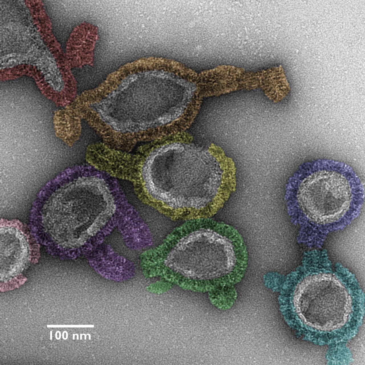
Description: The micrograph image provides insight into the assembly pathway of antimicrobial guanylate-binding proteins (GBP1s) at a high structural resolution. It adds to our understanding of GBP-orchestrated host immunity against bacterial infection. GPB1 forms an antimicrobial protein coat on bacteria and thereby plays a critical role in host defense against bacterial pathogens.
Method: A false-colored negative-stained transmission electron micrograph image shown at 73,000x nominal magnification, captured using a transmission electron microscope operating at 120 kV.
Watched over by vimentin's embrace

Description: The dynamics of vimentin protein fibers in endothelial cell monolayers under simulated blood flow conditions. Vimentin networks' show a morphological shift, aligning in the direction of the simulated flow, a phenomenon along with its nuclear “blanketing” effect that had not been observed before. Such flow-induced fiber alignment suggests a protective mechanism for cell nuclei.
Method: Captured with a Zeiss LSM 980 confocal microscope using a 63x 1.4NA oil objective in SR-4Y multiplex airy scan mode for super-resolution imaging, three separate excitation/emission filters and laser configurations were utilized to image different molecular targets. These were then overlaid using differently colored lookup tables. A total of 32 z-slices, 150 nanometers apart, were merged into a single image through Z-Projection. Color Lookup Tables (LUTs) were applied: Red for DAPI (DNA), MPI-Inferno for VE-Cadherin antibody, and Cyan Hot for vimentin antibody immunofluorescence, to distinguish the different elements clearly.
Text: Gunjan Sinha


