
Fibroblasts and mini organs: The most beautiful scientific pictures 2019
Every year at the Long Night of Science, visitors can decide: What is the most beautiful scientific picture of the year?
Dozens of researchers take part in the competition, which was initiated by Professor Helmut Kettenmann and his team. The pictures place scientific material in an artistic context and thus provide an unusual insight into research. The winners will receive a Nikon Coolpix camera, sponsored by the Society of Friends of the MDC and Nikon.
1st place
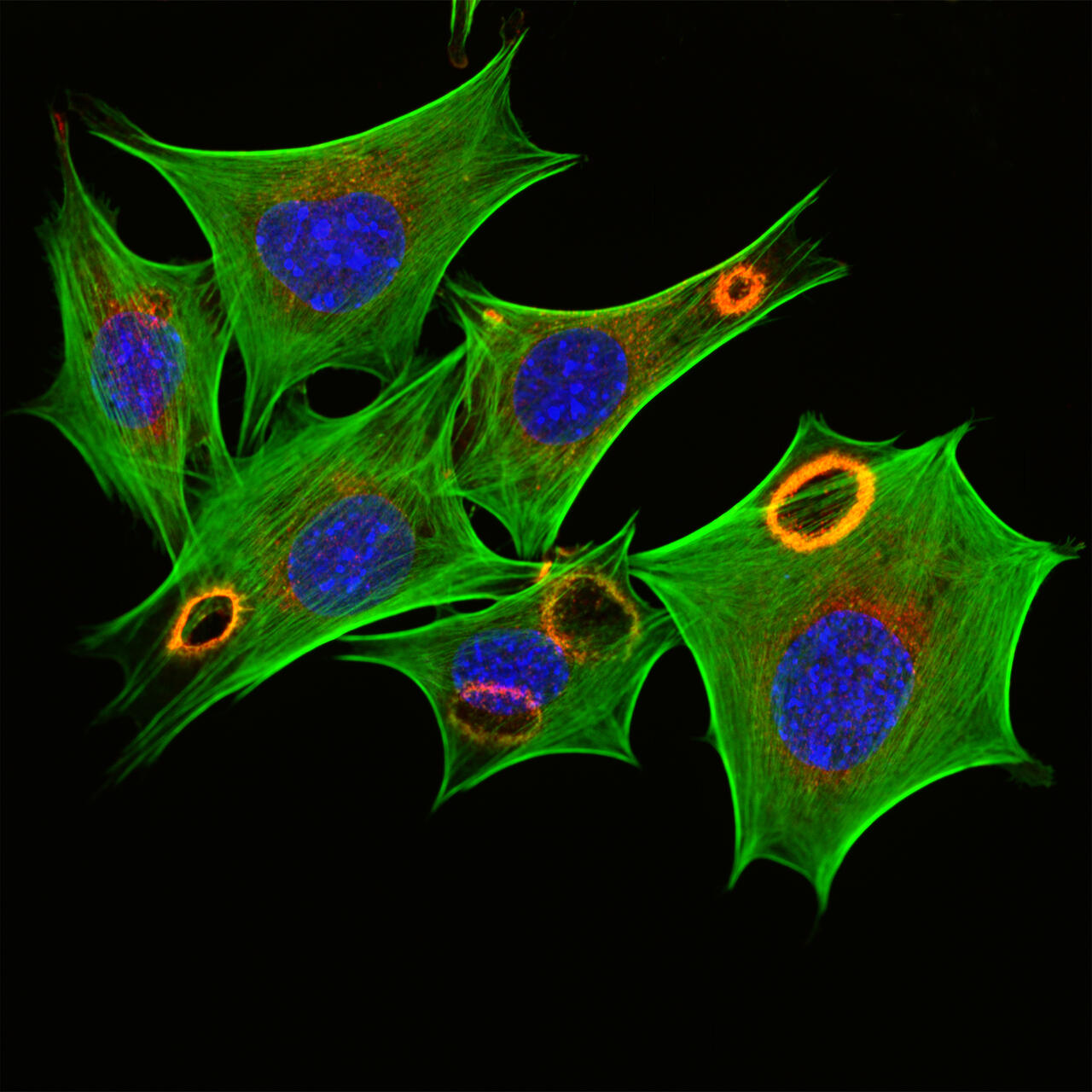
Fibroblasts, specific cells of the connective tissue stimulated with growth factors, can be seen. Growth factors are released in injuries from blood platelets, among other things, to stimulate tissue healing. Fibroblasts are stimulated to divide, migrate into wounds and produce collagen. Fluorescence staining shows the actin cytoskeleton (green), the cell nucleus (blue) and the protein cortactin (red). Cortactin is located at the dorsal (upper) cell membrane in ring-shaped vertical waves that spread like a fire and transform the actin cytoskeleton within a few minutes.
2nd place

Like us, the mouse has many receptors in its skin that recognize different sensory impressions, such as touch and temperature. The receptor TRPM8 recognizes menthol and is also significantly involved in the perception of cold. The receptors are marked with a fluorescent dye and are increasingly recognizable in the corkscrew-like structures of the sweat glands. These, in turn, are found mainly at the tips of the toe phalanges and the paw pads of the four-membered paw of the mouse. The illustration was created by a three-dimensional microscopic image and was color processed.
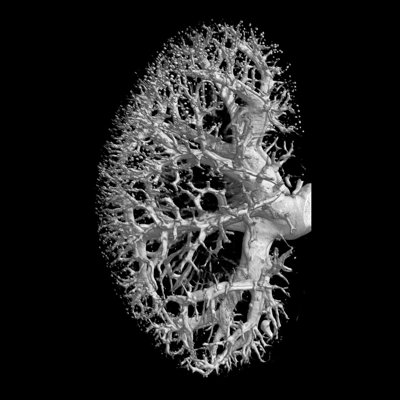
The picture shows the vessels of a mouse kidney. The kidney was imaged in 3D with a high-resolution computer tomograph (CT) with a resolution of 5 μm3 . Such detailed examinations enable the early detection of pathological vascular changes. Images taken in animals can make a decisive contribution to a better understanding of the causes of human kidney disease.
3rd place
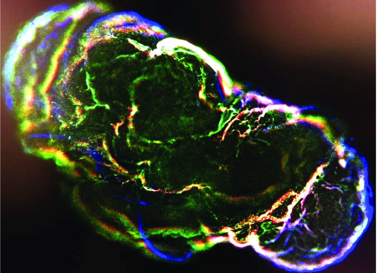
Three-dimensional miniorgans (organoids) can be produced from stem cells. In this case it is a composite heart-brain-organoid, which is supposed to imitate the control of the heart by the nervous system. Stem cells, which have developed into nerve cells, are coloured green, while the heart cells appear red (the cell nuclei in all cells are blue).


