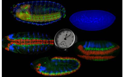
The beauty of science
Mini-organs are not only useful for cancer research; they also have certain aesthetic qualities – as one of the pictures in the Best Scientific Images Contest 2018 shows. This year, MDC scientists submitted 22 particularly impressive images from everyday laboratory life to the competition. The winners were selected during an exhibition held on the Long Night of the Sciences, where visitors could choose their personal favorites.
Every year, dozens of researchers take part in the competition – which was initiated by Professor Helmut Kettenmann and his team. The pictures frame scientific matter in an artistic context, thus providing an unusual insight into the work of the MDC. The winners each receive a Nikon Coolpix camera, sponsored by the Society of Friends of the MDC and Nikon.
Hannah Thurow
1st prize
A fruit fly embryo changes drastically in only about twelve hours:
Undifferentiated cells (2 o’clock) assume different identities (4 o’clock), migrate into individual positions (6‒10 o’clock) and produce differentiated cell types (12 o’clock), including nerve cells and muscle cells. The embryos are positioned with their heads to the left. 2 o’clock: cell nuclei (blue); 4‒10 o’clock: key genes for neuronal identity (RNA in situ hybridization, red, vnd; green, ind; blue, Dr); 12 o’clock: differentiated cell types (antibody coloring: red, cell bodies of neurons; green, axons of projection neurons; blue, muscle cells).
2nd prize
Is that a mini-pig in the Petri dish or a symbol of hope for colon cancer therapy?
This picture shows an intestinal organoid grown from mouse cells, with the stem cells stained green. When the Wnt signaling pathway is activated in these stem cells – as is the case in colon cancer patients – they multiply prolifically. By turning off certain genes, we can then acquire a molecular understanding of how colon cancer develops and find potential targets for therapy. The image was taken with a confocal laser scanning microscope belonging to the MDC’s Advanced Light Microscopy Platform. The cell edges are shown in red and the cell nuclei in blue.
3rd prize
This image shows a 15-day-old mouse embryo.
Organs such as the heart, liver, and brain are already largely developed. The umbilical cord clearly shows how the embryo is connected to its mother (large blood vessel with a strong red color seen bottom center). The structural protein actin is colored green, the protein EHD2 red, and the cell nuclei blue.


