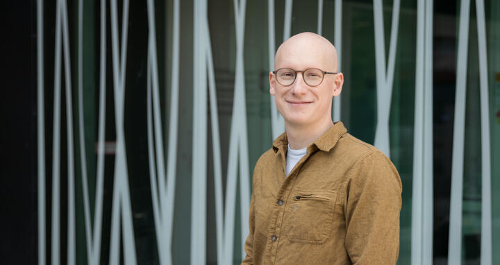
Unwinding clues to disease in DNA’s 3D structure
Dr Michael Robson has been contemplating the molecular origins of muscular dystrophies, a group of genetic diseases that cause progressive muscle loss, since his graduate student days at the University of Edinburgh. He later explored non-coding regions of DNA and their role in disease at Dr Stefan Mundlos’ lab at the Max Plank Institute for Molecular Genetics. "I've been considering this problem since my Ph.D. and I want to know what's actually going on," he says
The new Robson lab at the Berlin Institute of Medical Systems Biology at the Max Delbrück Center (MDC-BIMSB) in Berlin will study how disruptions in the three-dimensional structure of DNA can lead to genetic diseases such as muscular dystrophies.
A cell’s function is determined by the organization of DNA into tightly wound complexes of protein and DNA called chromatin. Proteins bind to the chromatin and loosen the packaging, making certain genes accessible for reading. Inaccessible genes are lodged at the envelope of the nucleus while the active ones move to the center. The Robson lab is interested in the interplay of molecules and events leading to genes to switch between these two states. Scientists think this process may play an as-yet-unknown role in the development of many genetic diseases such as muscular dystrophies and retinal disease.
“We don't really understand how all those pieces fit together,” Robson says. “By examining diseases where individual components are broken, we can determine how they should be normally function and can be repaired.”
What happens before a gene is switched on?
The Robson lab will be using a novel technology called ‘single cell Dam&T sequencing’: anytime a protein of interest attaches to DNA, a nearby segment gets labeled. This allows scientists to map the sequence of events leading to gene activation. The scientists have started mapping this process in healthy muscle cells and will study it further during the development of certain genetic diseases. They will be working with
mice and organoids, which can model of the human neuro-muscular system – but have their own limitations. “When you look in an actual organism, then suddenly you realize why all of these systems are so complicated,” Robson says.
“By doing studies like this, I want to see if we can explain to patients why they have this disease, and this mutation in this place has this effect,” he says. “At the same time, my personal fascination is just to understand how DNA controls genes.“
Text: gav
Further information
- Research at the muscular dystrophy at the Max Delbrück Center
- Neuromuscular organoids at the Max Delbrück Center


